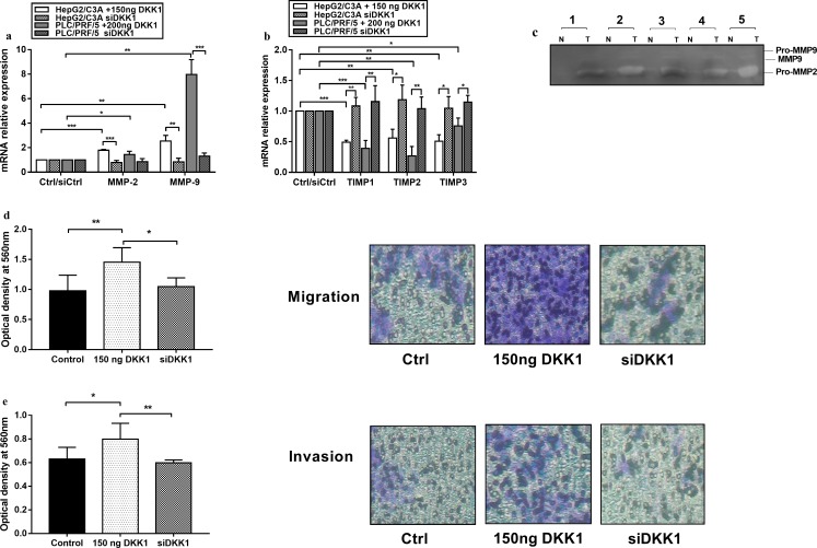
Fig 4. Recombinant DKK1 promotes the invasion and migration of hepatocellular carcinoma cell lines through shifting the MMPs/TIMPs ratio in favor of MMPs.
(a) Relative mRNA expression levels of MMP-2 and MMP-9 show an overall significant increase after PLC/PRF/5 and HepG2/C3A cells were exposed to DKK1 for 48 h and 72 h, respectively, but this was reversed when DKK1 was silenced. (b) Relative mRNA expression levels of TIMP-1, TIMP-2, and TIMP-3 showing an overall significant decrease after DKK1 treatment and an increase after DKK1 knockdown. (c) Zymography for detecting MMP activity in the supernatants from HepG2/C3A (lanes 1–3) and PLC/PRF/5 (lanes 4 and 5); T, treated with 150 ng/ml and 200 ng/ml recombinant DKK1, respectively; N, untreated cancer cells. An obvious increase occurred in the latent form of MMP-2 in response to DKK1. Both the latent and active forms of MMP-9 were observed in the treated cells. (d) Migration assay of HepG2/C3A by Transwell Boyden chamber in the presence of 150 ng/ml recombinant DKK1 and in the absence of DKK1 expression. Migratory cells on the bottom of the polycarbonate membrane were stained and quantified based on the OD at 560 nm. Representative photographs of migratory HepG2. (e) Invasion of HepG2/C3A cells treated with 150 ng/ml recombinant DKK1, and DKK1 knock down cells assessed by the Transwell Boyden chamber. Invasive cells on the bottom of the invasion membrane were stained and quantified based on the OD at 560 nm. Representative photographs of invasive HepG2. The data are representative of at least 3 independent experiments and are expressed as the mean ± SD. * p <0.05, ** p <0.01, *** p <0.001, as indicated.

