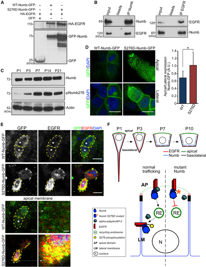
Figure 4. EGFR Trafficking via Numb Phosphorylation.
(A) IP from HEK293 cell lysates co-transfected with constructs as indicated (+) and probed with antibodies to HA (EGFR, upper blot) and GFP (Numb, lower blot).
(B) IP from P3 LV whole mounts using anti-Numb or anti-EGFR antibodies. Blots were then probed with anti-Numb and anti-EGFR antibodies.
(C) Western blot analyses of LV walls of indicated ages and blotted for Numb and pNumb276. Actin is the loading control.
(D) IHC images of MDCK cells expressing either WT-Numb-GFP or S276D-Numb-GFP stained with anti-GFP antibody and DAPI. Single optical section views are from cellular apical surfaces (apical view, top row) or below the surface showing lateral membrane domains (lateral view, bottom row). Scale bars: 10 mm. The ratio of GFP fluorescent intensity at apical versus lateral domains for WT-Numb-GFP and S276D-Numb-GFP is shown. *p < 0.001, Student’s t test, n = 10, mean ± SEM.
(E) IHC images of P7 LV whole mounts expressing WT-Numb-GFP or S276D-Numb-GFP (from P1 lentiviral infection) labeled with anti-GFP, EGFR antibodies, and DAPI. Note high-level EGFR expression in S276D-Numb-GFP-expressing cells, but not WT-Numb-GFP-expressing cells (*). Right panels: single optical plane of the apical membrane, with enlarged areas showing high-level EGFR localization with S276D-Numb-GFP (dashed boxes). Scale bars: 10 µm; inset 1 µm.
(F) Schematic illustrations showing EGFR/Numb localizations during postnatal ependymal maturation (top) and the putative molecular pathway of EGFR redistribution (bottom).

