Figure 2.
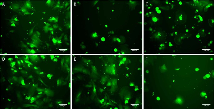
Cell spreading assay to CMPs. A–E, morphology of hMSCs (green) after 4 h in culture adhered to collagen III (positive control) (A), BSA (negative control) (B), GROGER peptide (negative control) (C), T3-237 WT (D), G240A (E), and G240V (F). Cell attachment and morphology on the WT and G240A match that on the positive control, whereas attachment and morphology on G240V are similar to that on the negative controls.

