Figure 2.
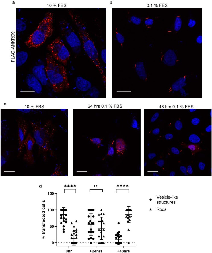
ANKRD9 forms rods upon nutrient depletion. a, HeLa cells were transfected with FLAG-ANKRD9 (red) as in Fig. 1a and incubated in the presence of 10% FBS. n = 3 independent experiments. b, HeLa cells were transfected with FLAG-ANKRD9 and incubated in the presence of 0.1% FBS for 48 h. Rods are indicated by gray arrows; nuclei are stained with DAPI (blue). n = 3 independent experiments. c, representative images of each time point; rods appear in the majority of cells after 48 h in 0.1% FBS media. For 24 h, n = 3. Scale bar, 20 μm. d, percentage of cells containing ANKRD9 in vesicle-like structures (black filled circles) or rods (black filled triangles) after overnight transfection (control), followed by 24 or 48 h in starvation (0.1% FBS) media. The total number of analyzed cells is as follows: n = 137 cells (control, 0 h of 0.1% FBS), n = 149 cells (24 h of 0.1% FBS), n = 131 cells (48 h of 0.1% FBS). Error bars represent S.D. Each individual point represents at least 5 cells. ****, p value < 0.0001., unpaired t test; ns, non-significant.

