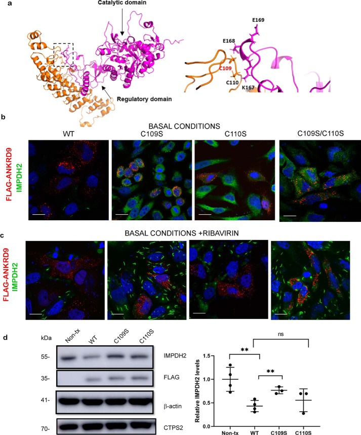
Figure 7.
The Cys109Cys110 motif contributes to ANKRD9-IMPDH2 interaction. a, left, docking model of ANKRD9 and IMPDH2 (PDB ID 1NF7, see Fig. S7). IMPDH2 catalytic and regulatory domains are indicated by the arrows; the predicted interaction site is indicated by the box. Right, the magnified view of the predicted interaction site between ANKRD9 (orange) and IMPDH2 (purple). The CysCys motif in ANKRD9 interacts with Lys167, Glu168 and Glu169 of IMPDH2. b, Cys mutants do not facilitate IMPDH2 degradation. HeLa cells were transfected with WT ANKRD9 and indicated mutants under basal conditions (10% FBS) and immunostained for FLAG and IMPDH2. Cells expressing WT ANKRD9 showed significantly reduced IMPDH2 staining whereas cells with the Cys mutants did not. n = 3 independent experiments for each condition. Scale bar, 20 μm. c, Cys mutants of ANKRD9 do not form rods with IMPDH2. HeLa cells were transfected with the indicated constructs and subsequently treated with 10 μm ribavirin for 1 h prior to immunostaining for FLAG and IMPDH2. n = 3. Scale bar is 20 μm. d, left, the WT and ANKRD9 mutants were expressed in HeLa cells under basal conditions (10% FBS) and cell lysates, were separated and immunoblotted for FLAG, IMPDH2, CTPS2, and β-actin used as a loading control. WT ANKRD9 but not the C109S mutant significantly decreases IMPDH2 abundance, the C110S mutant is intermediate between the two and similar to WT. Right, densitometry of IMPDH2 intensity after WT ANKRD9 or mutant expression in HeLa cells. n = 3. ****, p value < 0.0001., unpaired t test.

