Figure 3.
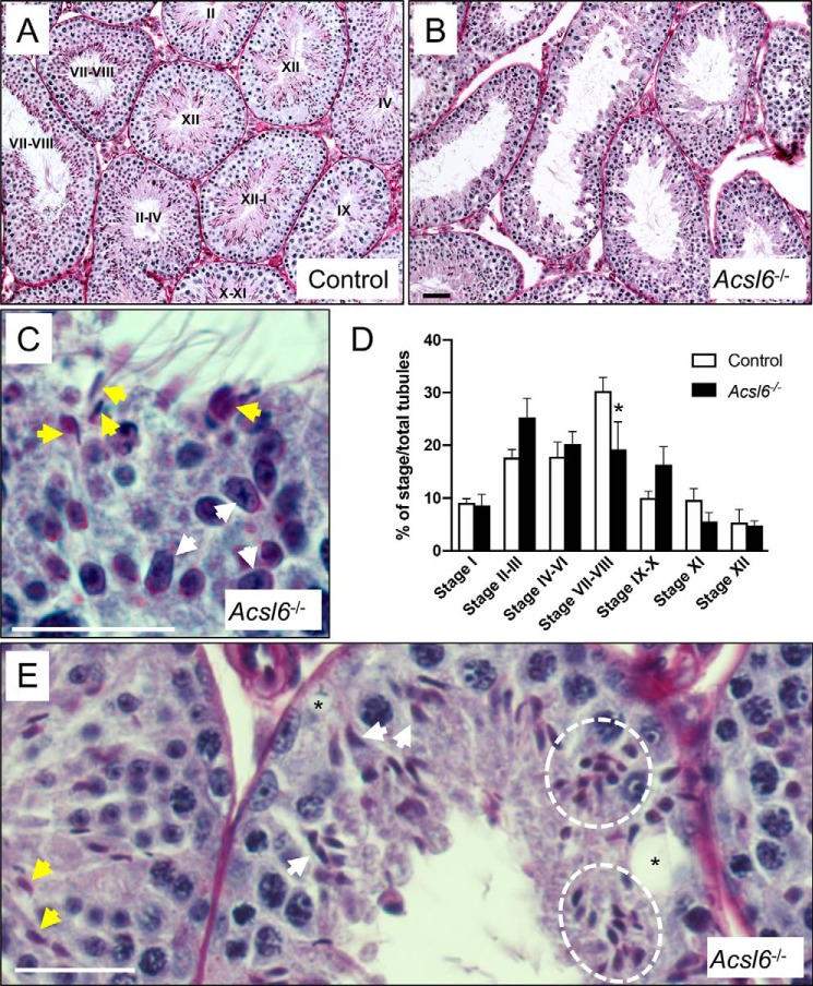
ACSL6 deficiency disrupts spermatogenesis. A and B, testis sections from control (A) and Acsl6−/− (B) mice were PAS-stained. Stages are indicated in the lumina of control tubules. C and D, sections from PAS-stained Acsl6−/− testes are enlarged to show detail. C, a tubule at stage IX (because of early elongating spermatids, white arrows) also erroneously contains condensed spermatids that should have already been released at spermiation (yellow arrows). D, seminiferous epithelial stages were quantified in control and Acsl6−/− testes, and there was a significant decline in the numbers of stages VII–VIII. E, tubules contain normal-appearing condensing spermatids (yellow arrows), as well as individual (white arrows), and clusters (white dashed circle) of misshapen spermatid heads, as well as vacuoles (asterisks). The data represent averages ± S.E., and an asterisk indicates significance at p ≤ 0.05 by Student's t test. Scale bars in B–D, 60 μm.

