Figure 3.
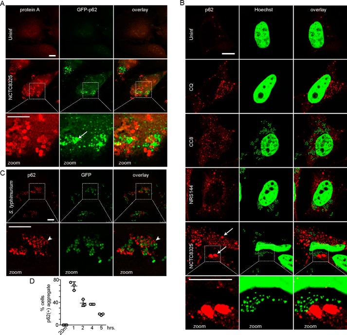
Differential formation of p62/Sequestosome1-positive membranes following infection by Staphylococcus versus Salmonella. A, HeLa/GFP–p62 cells were infected with 100 m.o.i. NCTC8325 for 3 h (gentamicin added after 1st h). After fixation, bacteria were detected by anti-protein A staining. Arrow, p62(+) aggregate. All scale bars, 10 μm. All zoom: ×3.4 magnification. B, HeLa cells were treated with chloroquine as control (CQ, 25 μm) or infected as above with indicated strains of S. aureus. After fixation, cells were stained with antibodies for p62/SQSTM1 and Hoechst 33342 (detects bacterial and host cell DNA). Arrows: large size p62(+) aggregates. C, HeLa cells were infected with 1:100 diluted GFP–S. enterica sv. typhimurium for 1 h before fixation and staining with antibodies for p62/SQSTM1. Arrowhead, co-localization of p62 on Salmonella. D, HeLa cells were infected with GFP–Salmonella as in C for the indicated times before fixation. The percentage of cells positive for p62(+) membranes was counted (40–110 cells counted per sample). Average from n = 3 samples ± S.D. Uninf., uninfected.

