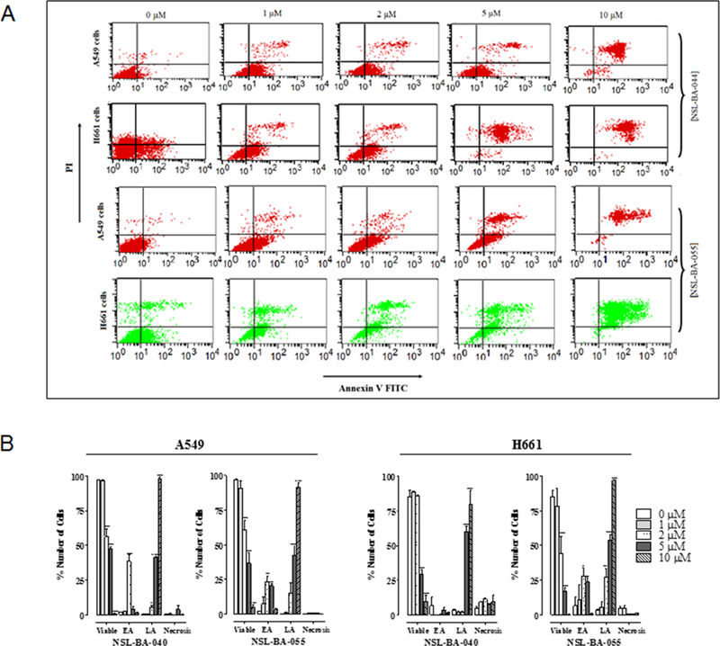
Figure 4. PCAIs induce apoptosis.
A549 and H661 cells treated with PCAIs (1 – 10 μM) for 48 h were examined for externalization of phophatidylserine using Annexin V-FITC and flow cytometry. PCAI treatment promoted an increase in the number of cells with an increased Annexin V-FITC fluorescence prior to the loss of membrane integrity in early apoptosis (lower right-hand quadrant) and late apoptosis (upper right-hand quadrant). The dot plots (A) represent a single experiment while the graph (B) is an average from 3 independent experiments.

