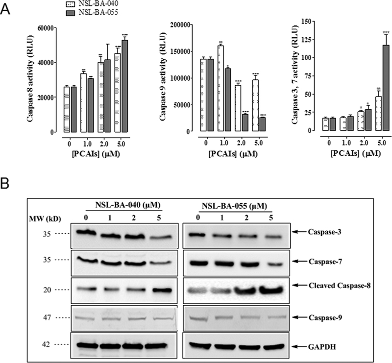
Figure 5. PCAIs-induced apoptosis is mediated by Caspase-8 and Caspase-3/7.
(A) A549 cells were seeded at 5000/well for Caspase-3/7 assay and 30,000/well for both Caspase-8 and 9 assays in a white-walled 96-well plate for 24 h before start of experiment. PCAIs (1 −5 μM) or 1μL of acetone (carrier solution) were then added to triplicate wells. Identical amounts of PCAIs were used to supplement the samples at 24 h for 48 h exposure. Caspase activity was determined after 48 h incubation with Caspase-glo luminescent assay according to manufacturer’s instructions. Data on relative luminescence units (RLU) are expressed as mean ± S.D. Significance (*p < 0.05, **p < 0.01, ***p < 0.001) was determined by Student’s t-test. (B) A549 cells (2×10⁵ cells/well) grown in 100 mm tissue culture dishes were treated with PCAIs (0 – 5 μM) for 48 h. Cell lysates were generated using RIPA lysis buffer, protein concentration was determined and lysates containing equal amounts of proteins were separated by gel electrophoresis and proteins transferred onto polyvinylidene difluoride (PVDF) membranes. Membrane-bound proteins were probed with antibodies against caspase-8, caspase-9, caspase-3/7, GAPDH and visualized using HRP-conjugated secondary antibodies and ECL reagents.

