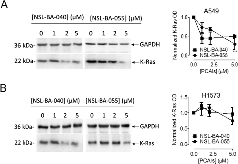
Figure 6. PCAIs diminish K-Ras protein levels in lung cancer cells.
Whole cell lysates of lung cancer cells, A549 and H1573 treated for 24 h with PCAIs (0 −5 μM) were analyzed by western blot for K-Ras protein levels expression as described in the Materials and Methods. Briefly, 40–50 μg of whole cell lysate proteins were loaded into the wells of 12% gels and subjected to SDS-PAGE electrophoresis. Proteins were then transferred unto PVDF membranes and membranes were immunoblotted for K-Ras and GAPDH. Densitometry of bands was performed using Image Lab Software and normalized to GAPDH. Data are representative of three independent experiments. Statistical significance (**p < 0.01) was determined by 1-way ANOVA with post hoc Dunnett’s test.

