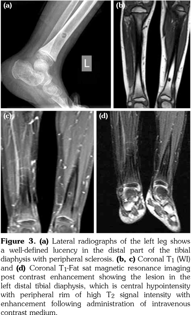
Figure 3. (a) Lateral radiographs of the left leg shows a well-defined lucency in the distal part of the tibial diaphysis with peripheral sclerosis. (b, c) Coronal T<sub>1</sub> (WI) and (d) Coronal T<sub>1</sub>-Fat sat magnetic resonance imaging post contrast enhancement showing the lesion in the left distal tibial diaphysis, which is central hypointensity with peripheral rim of high T<sub>2</sub> signal intensity with enhancement following administration of intravenous contrast medium.

