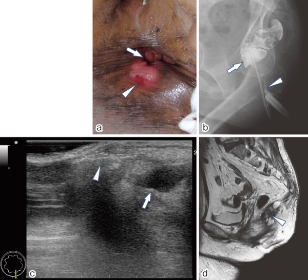
Figure 1.

Physical and radiologic images.
(a) Physical examination shows a small, accessory opening (arrow head), located at midline, approximately 1 cm posterior to the true anus (arrow).
(b) Fistulography shows a 2-cm canal (arrow head) without connecting with the rectum (arrow).
(c) Endoanal ultrasonography shows a brightly hyperechoic blind-ending fistulous tract (arrow head) and a hypoechoic cystic mass (arrow) in the superior retrorectal area.
(d) Magnetic resonance imaging shows a multiloculated presacral cyst (arrow head) posterior to the rectum.
