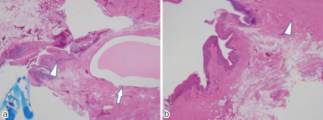
Figure 3.
(a) The duct is predominantly lined by squamous epithelium (arrow head) and the cyst is lined by squamous, columnar, and transitional epithelia (arrow). (hematoxylin-eosin stain, 40×).
(b) Smooth muscle bundles are present in the wall of the duct (arrow head). (hematoxylin-eosin stain, 40×).

