
Fig. 1.
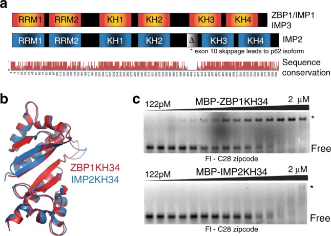
ZBP1 and IMP2 overview and crystal structures. a Overall domain structure and sequence conservation of the IMP family members above. Below, sequence conservation across the IMP family members. b Ribbon diagrams overlaying the crystal structures of ZBP1KH34 in red (PDB: 3KRM) and IMP2KH34 in blue (this study, PDB:6ROL). c Representative EMSAs for wild-type C28 ß-actin zipcode RNA. The filled triangle represents a 1:1 serial dilution of ZBP1KH34 (top) and IMP2KH34 (bottom). The RBP–RNA complex (*) and free RNA (FREE) are labeled