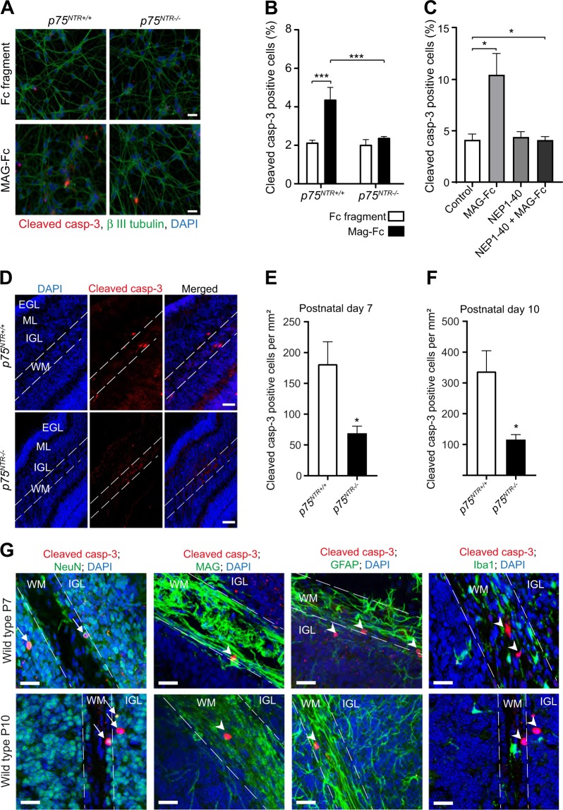
Fig. 3. MAG induces cell death in developing CGNs.
a Representative image of P7 p75NTR+/+ and p75NTR−/− CGNs cultured for 2 days in vitro prior to treatment with either Fc fragment (control, 25 μg/ml) or MAG-Fc (25 μg/ml) for 24 h. After treatment, cells were stained with anti-cleaved casp-3 (red), anti-β III tubulin (green) and counterstained with DAPI (blue). Scale bars, 20 μm. b Percentage of cleaved casp-3 positive neurons in cultured p75NTR+/+ and p75NTR−/− CGNs treated with Fc fragment (control, 25 μg/ml) or MAG-Fc (25μg/ml) for 24 h (150 images per genotype and condition). Mean ± s.e.m. of data from four separate cultures, ***p < 0.001 compared to control (two-way ANOVA followed by Bonferroni post hoc test). c Percentage of cleaved casp-3 positive neurons in cultured wild type CGNs treated with either MAG-Fc (25 μg/ml) alone, NgR antagonist (NEP1-40; 10 μM) alone or MAG-FC plus NEP1-40 for 24 h (40 images per genotype and condition). Mean ± s.e.m. of data from three separate cultures, *p < 0.05 compared to control (one-way ANOVA followed by Bonferroni post hoc test). d Representative images of folium III of P7 cerebellum double stained with anti-cleaved casp-3 and DAPI. The outline shows the white matter in folium III. Scale bars, 50 μm. e–f Quantification of cleaved casp-3 positive cells in the white matter of folium III from P7 (e) and P10 (f) p75NTR+/+ and p75NTR−/−cerebella. Mean ± s.e.m, n = 6 mice per genotype; *p < 0.05; unpaired Student’s t test. g Micrographs of folium III of P7 cerebellar sections immunostained for cleaved casp-3 (red) together with either anti-NeuN (green), anti-MAG (green), anti-GFAP (green) or anti-Iba1 (green) and counterstained with DAPI (blue). Arrows shows neurons double-positive for cleaved casp-3 and NeuN while arrow heads indicates neurons positive for cleaved casp-3 that are negative for MAG, GFAP or Iba1. The outlines show the white matter. Scale bars, 50 μm

