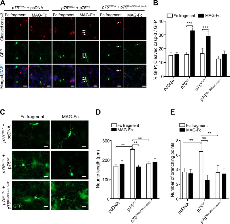
Fig. 6. MAG-induced cell death requires coupling of p75NTR to RhoGDI/cell death pathway.
a, b Representative images (a) and quantification (b) of P7 p75NTR−/− CGNs transfected on the second day in vitro with either pcDNA (vector), p75WT or p75RhoGDI/cell death constructs. Twenty-four hours after transfection, neurons were treated with either 25 μg/ml Fc fragment or 25 μg/ml MAG-Fc for 24 h and triple-labelled with anti-cleaved casp-3, anti-GFP and DAPI. Arrows indicated neurons double-positive for GFP and cleaved casp-3. Scale bars, 20 μm. The percentage of cleaved casp-3 positive P7 p75NTR−/− CGNs (b) is shown as mean ± s.e.m. of data from four separate cultures, total of 60 images per genotype and condition (***p < 0.001 compared to control, two-way ANOVA followed by Bonferroni post hoc test). c–e Representative images (c) and quantification (d, e) of P7 p75NTR−/− CGNs transfected on the second day in vitro with either pcDNA (vector) or p75WT or p75RhoGDI/cell death constructs and treated 24 h after transfection with either 25 μg/ml Fc fragment or 25 μg/ml MAG-Fc for 24 h. The neurons were labelled with anti-GFP. Scale bars, 20 μm. The mean length of the longest neurite (d) and the branch point number (e) are shown as mean ± s.e.m. of data from three separate cultures, total of 60 images per genotype and condition (**p < 0.01, compared to control, two-way ANOVA followed by Bonferroni post hoc test)

