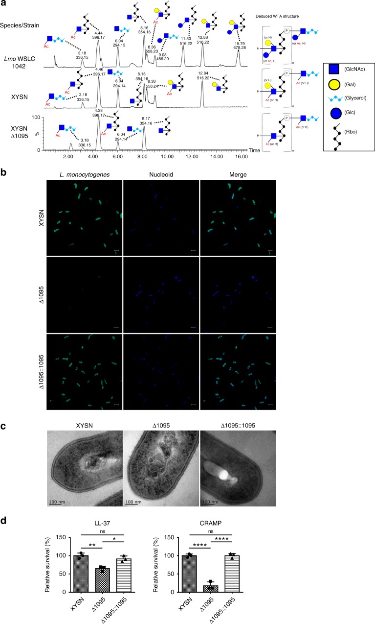
Fig. 5.
Molecular and structural features of wall teichoic acid of L. monocytogenes HSL-II strain XYSN. a Structure and UPLC-MS/MS analysis of type II WTA from L. monocytogenes WSLC 1042 serotype 4b strain as compared with XYSN and its isogenic Δ1095 mutant. Liquid chromatographic separation and MS-based identification of carbohydrate residues within Listeria WTAs from XYSN, Δ1095, and WSLC 1042. Peaks are labeled with their respective retention time (Rt;[min]) and base peak ion [M-H]− (m/z). b Confocal images of XYSN, Δ1095, and Δ1095::1095. Bacteria were subsequently stained by Listeria O-antiserum 4 (primary antibody, 1:200) and Alexa Fluor 488-conjugated rabbit antibody (green), nucleoid was stained by DAPI (blue). Magnification of all images: ×3000. Scale bars, 2 μm. c Transmission electron microscopic images depicting intact cell wall structures of XYSN, rough cell wall structure of Δ1095 and regained intact cell wall of Δ1095::1095. d Role of galactosylated WTA in protection of bacteria towards antimicrobial peptides. Quantification of viable bacteria following treatment (2 h 37 °C) with LL-37 and CRAMP antimicrobial peptide (25 µg/mL each) on growing cultures. Statistical analyses were carried out by Tukey's multiple comparisons test: *P < 0.05, **P < 0.01, ****P < 0.0001, ns: no significance. Error bars represent SD, n = 3 independent experiments

