Fig. 1.
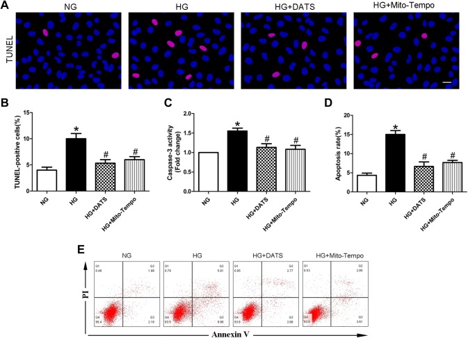
Effect of DATS on inhibition of high glucose-induced HUVEC apoptosis in vitro. a TUNEL assay. HUVECs were seeded, treated with different agents, then grown under normal glucose (NG) or high-glucose (HG) conditions, and subjected to the TUNEL assay. The images show DAPI-stained (blue) and TUNEL-positive cells (pink); scale bar = 25 μm. b Graph shows the quantified HUVEC apoptosis data. c Caspase-3 activity assay. HUVECs were seeded, treated with different agents, then grown under normal glucose (NG) or high-glucose (HG) conditions, and subjected to the caspase-3 activity assay. d Graph shows the quantified HUVEC apoptosis data determined by flow cytometry. e Apoptosis was determined by flow cytometry. HUVECs were seeded, treated with different agents, grown under normal glucose (NG) or high-glucose (HG) conditions, and subjected to annexin V/PI staining for flow cytometry detection. The data are expressed as mean ± SEM (n = 3). *p < 0.05 versus the NG group and #p < 0.05 versus the HG group

