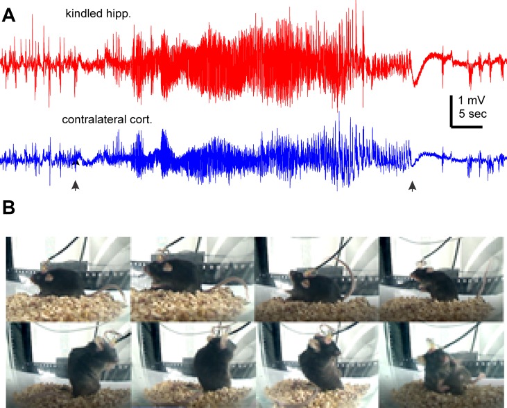
Figure 1.
Representative EEG discharges and corresponding motor seizure. EEG traces and images were collected from a mouse the first day after termination of kindling stimulation. (A) Ictal discharges were recorded from of the kindled hippocampus and contralateral parietal cortex. Filled arrows denote the onset and tarnation of discharges. (B) Sequential images (from top-left to bottom right) show a stage-5 seizure.

