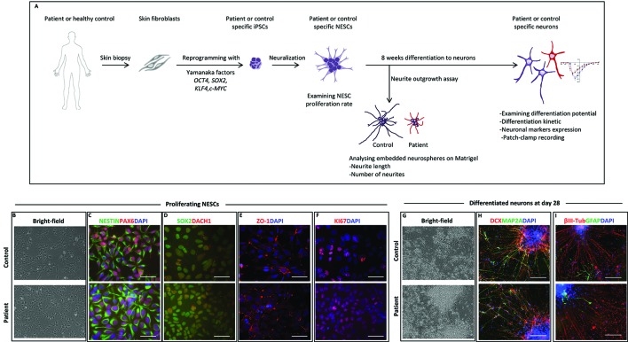
Figure 2.
Neural stem cell (NESC) characterization and differentiation. (A) Schematic diagram of our in vitro model from somatic cells to neurons. (B) Bright-field images of iPSC-derived NESCs from control and patient (III:4) in monolayer culture. NESCs self-organized into neural rosette structures. (C) Immunofluorescent staining of iPSC-derived NESCs. All derived NESC lines expressed neural stem cell markers NESTIN, PAX6, (D) SOX2, DACH1, (E) ZO-1, and (F) proliferation marker KI67. Nuclei stained with DAPI, scale bar: 50 µm. (G) Bright-field images of 28 days’ differentiated neurons of patient and control. (H) Immunofluorescent staining of 28 days’ differentiated neurons from patient and control. Differentiated neurons expressed neuronal markers DCX and MAP2A. (I) The majority of 28 days’ differentiated cells were positive for the neuronal marker ßIII-tubulin with low amount of cell expressing the glia marker GFAP. Scale bar 100 µm.

