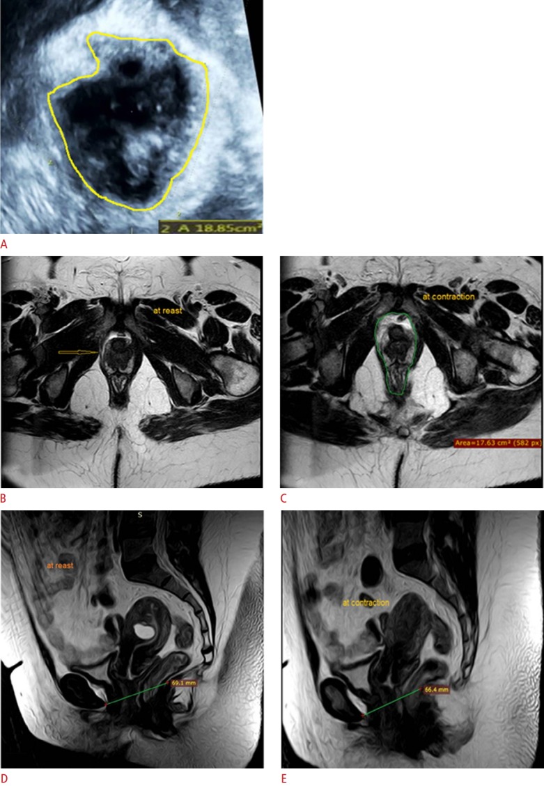
Fig. 3. A 44-year-old multipara (4 births) woman with a pelvic lump, urinary incontinence, and dyspareunia.

Digital palpation revealed rectocele and cystocele with no avulsion and a Modified Oxford Score grade of 3. A. Three-dimensional transperineal ultrasonography at contraction reveals avulsion (arrow) of the right puborectalis muscle (hiatal area=18.85 cm2 ). B, C. Magnetic resonance imaging (MRI) axial cuts show complete avulsion of the right puborectalis muscle (arrow in B) with thinning and fatty changes at rest (B) and contraction (C). D, E. MRI sagittal cuts show a reduction in the levator hiatal antero-posterior diameter from 69.1 mm at rest (D) to 66.4 mm at contraction (E).
