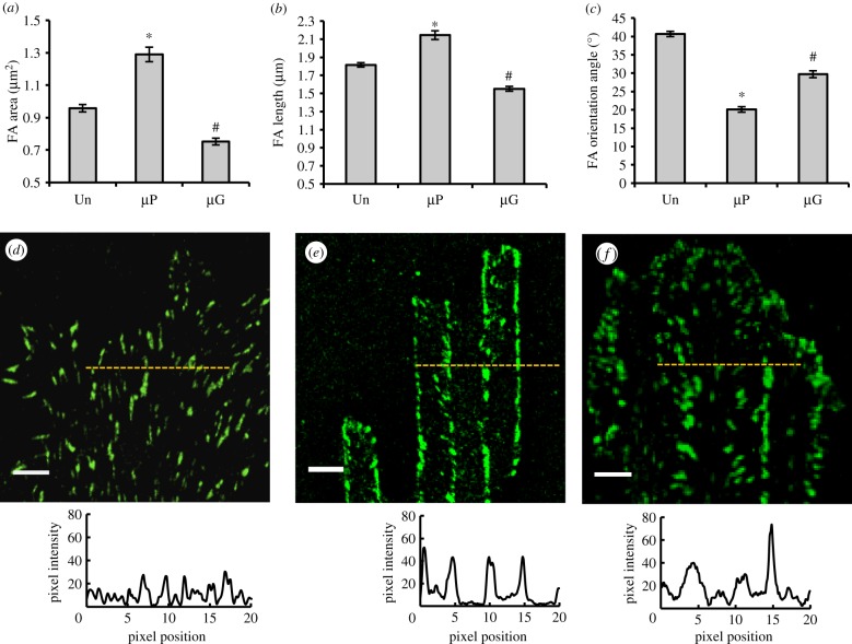
Figure 4.
Focal adhesion (FA) maturation on control and patterned surfaces: FA area (a), length (b) and orientation (c) of BAECs cultured for 24 h on pattered and control surfaces. 1296, 876 and 668 FAs from three independent experiments were measured on unpatterned, µP and µG substrates, respectively. Data are mean ± s.e.m. Asterisks (*) indicate statistically significant differences relative to the corresponding Un case (p < 0.01), while hashtags (#) denote statistically significant differences relative to both the Un and µP cases (p < 0.01). Confocal images of vinculin (green) and corresponding fluorescence intensity distribution profiles of BAECs cultured for 24 h on unpatterned (d), µP (e) and µG (f) substrates. The zero position corresponds to the beginning of an adhesive stripe for the planar bio-adhesive patterned surface and the beginning of a ridge for the microgrooved patterned surface. Scale bar is 5 µm. (Online version in colour.)

