. 2019 Sep 26;34(3):291–301. doi: 10.3803/EnM.2019.34.3.291

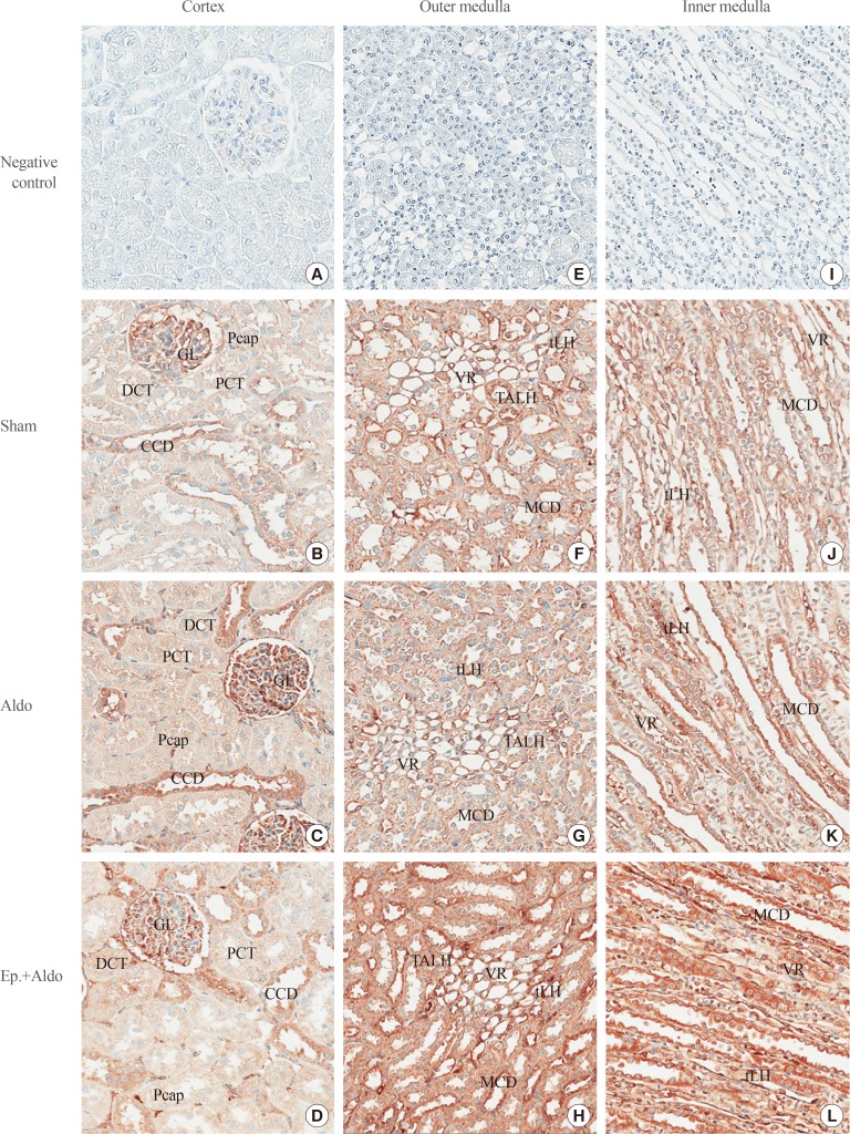
Fig. 2. Effects of aldosterone on renal striatin localization. Representative immunohistochemical staining micrographs of renal striatin in the cortex (A–D), the outer medulla (E–H), and the inner medulla (I–L) from the sham (B, F, J), aldosterone (Aldo) (C, G, K), and eplerenone and aldosterone (Ep.+Aldo) (D, H, L) groups (n=6/group). Negative controls: A, E, I (A–D, ×400; E–L, ×200). GL, glomerulus; Pcap, peritubular capillary; DCT, distal convoluted tubule; PCT, proximal convoluted tubule; CCD, cortical collecting duct; tLH, thin limb of the loop of Henle; VR, vasa recta; TALH, thick ascending limb of the loop of Henle; MCD, medullary collecting duct.

Fig. 2