. 2019 Sep 26;34(3):291–301. doi: 10.3803/EnM.2019.34.3.291

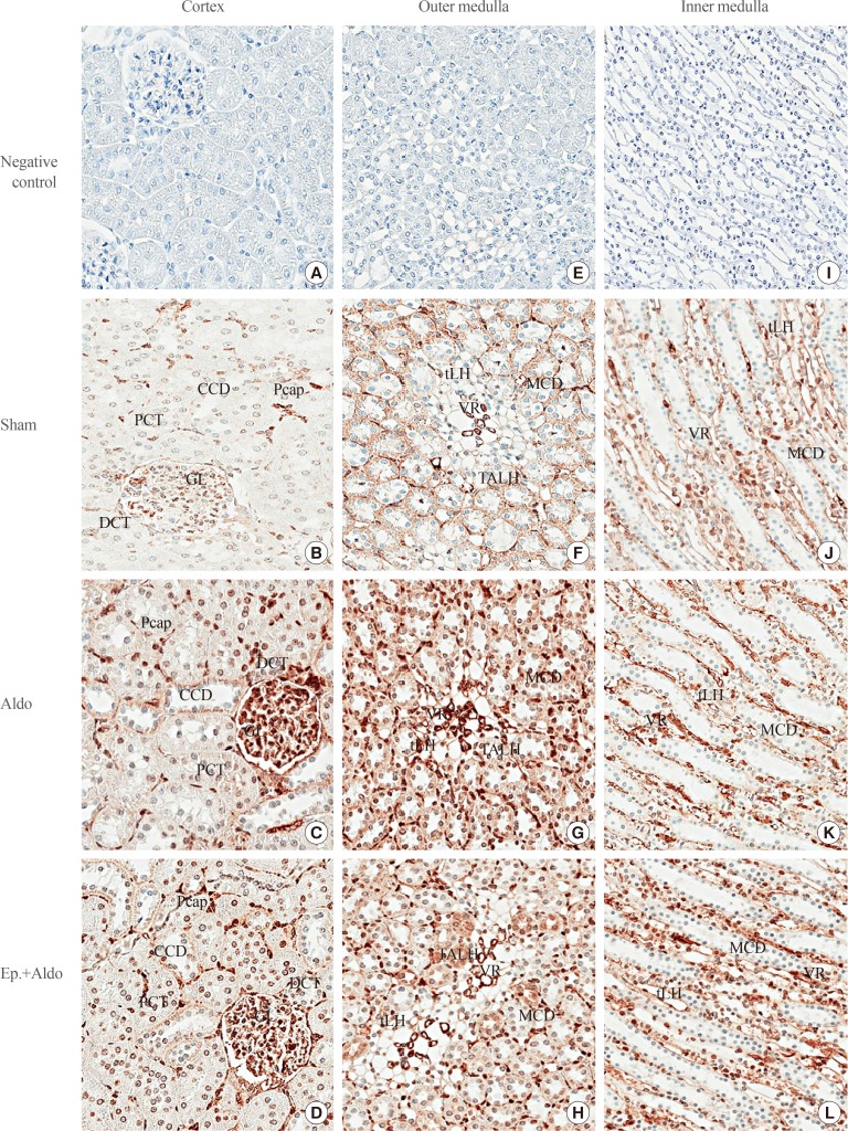
Fig. 3. Effects of aldosterone on renal cav-1 localization. Representative immunohistochemical staining micrographs of renal cav-1 in the cortex (A–D), the outer medulla (E–H), and the inner medulla (I–L) from the sham (B, F, J), aldosterone (Aldo) (C, G, K), and eplerenone and aldosterone (Ep.+Aldo) (D, H, L) groups (n=6/group). Negative controls: A, E, I (A–D, ×400; E–L, ×200). CCD, cortical collecting duct; Pcap, peritubular capillary; PCT, proximal convoluted tubule; GL, glomerulus; DCT, distal convoluted tubule; tLH, thin limb of the loop of Henle; MCD, medullary collecting duct; VR, vasa recta; TALH, thick ascending limb of the loop of Henle.

Fig. 3