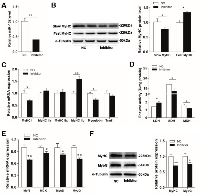
Figure 3.
The effects of miR-152 inhibitor transfection on myofiber specification and skeletal myogenesis in porcine myotubes. (A) The miR-152 level was examined by RT-PCR at four days post transfection. (B) The protein expression of MyHC isoforms in porcine myotubes was detected at four days after transfection with the miR-152 inhibitor. α-Tubulin served as an internal control. (C) MyHC isoforms and oxidative fiber markers in porcine myotubes transfected with the miR-152 inhibitor were determined by RT-PCR. (D) Metabolic-related enzymes activities in porcine myotubes transfected with the miR-152 inhibitor. (E) Relative mRNA levels of Myf5, MCK, MyoD, and MyoG were detected by RT-qPCR at four days after miR-152 inhibitor transfection. (F) The protein expression of myogenic markers, MyHC and MyoG, were detected by western blot at four days after miR-152 inhibitor transfection and analyzed using image J. Results are exhibited as mean ± S.E.M. (n = 4, four independent experiments). * p < 0.05, ** p < 0.01 and *** p < 0.001 when contrasted with the control.

