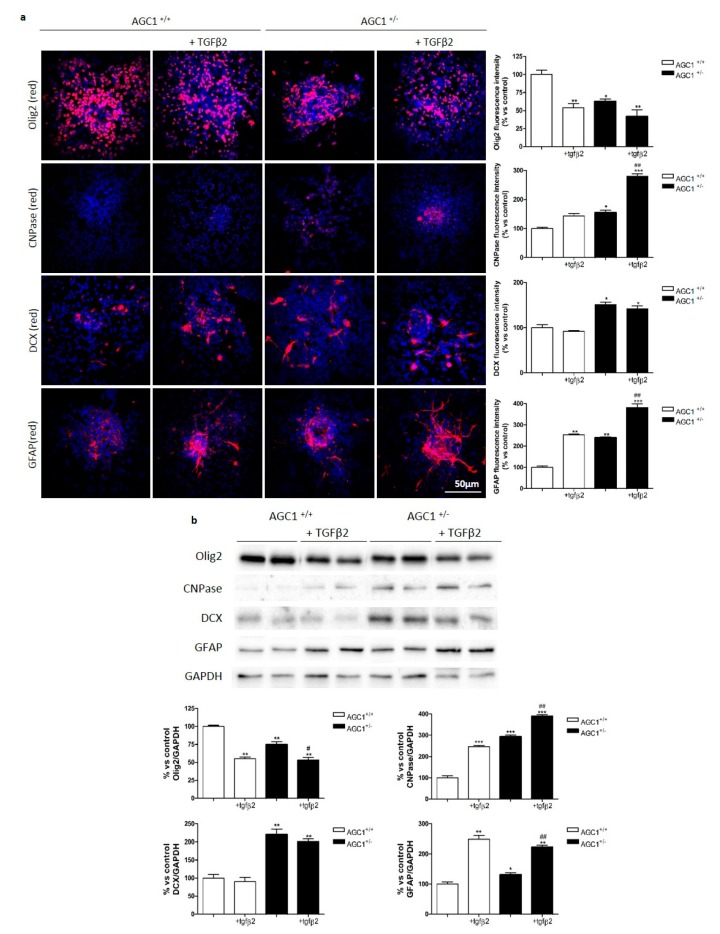
Figure 7.
Effect of exogenous TGFβ2 on proliferation/differentiation in neurospheres from AGC1+/+ and AGC1+/− mouse SVZ. Immunofluorescence staining in AGC1+/+ and AGC1+/− neurospheres and following treatment with TGFβ2 after 4-day differentiation (a). Olig2, CNPase, DCX and GFAP used as specific markers for OPCs, mature oligodendrocytes, NSCs and astrocytes respectively (nuclei labelled with Hoechst). Scale bar: 50 μm. Analyses for Olig2+ and DCX+ cell number and CNPase+/GFAP+ fluorescence signal intensity evaluated with Fiji ImageJ2 software. Values are the mean ± SE of 3 independent experiments performed in triplicate, * p < 0.05, ** p < 0.01, *** p < 0.001 compared to control AGC1+/+ neurospheres, # p < 0.05, ## p < 0.01 compared to control AGC1+/− neurospheres, Two-way ANOVA (Bonferroni’s post-test). Western blot analysis and relative densitometries of Olig2, CNPase, DCX and GFAP expression in AGC1+/+ and AGC1+/− neurospheres and following treatment with TGFβ2 after 4-day differentiation (b). Densitometry is the ratio between the expression level of each protein and GAPDH as reference loading control and is expressed as percentage vs AGC1+/+ neurospheres. Values are the mean ± SE of 3 independent experiments performed in duplicate, * p < 0.05, ** p < 0.01, *** p < 0.001compared to AGC1+/+ neurospheres, # p < 0.05, ## p < 0.01 compared to control AGC1+/− neurospheres, Two-way ANOVA (Bonferroni’s post-test).

