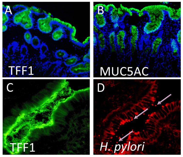
Figure 1.
Trefoil factor 1 (TFF1), the mucin MUC5AC, and Helicobacter pylori are found at the same sites in the human stomach. Formalin-fixed gastric mucosal tissue from H. pylori-negative individuals were immunofluorescently stained using specific antibodies against (A) TFF1 and (B) MUC5AC. Both TFF1 (green) and MUC5AC (green) staining occurred on gastric surface foveolar cells and in the glands. Cell nuclei were counter stained with DAPI (blue). Original magnification 100×. A frozen section of H. pylori infected antral gastric biopsy tissue was immunofluorescently stained using specific antibodies against (C) TFF1 and (D) H. pylori. Both TFF1 (green) and H. pylori (red) were detected at the epithelial surface and in the overlying gastric mucus. Pink arrows indicate H. pylori organisms staining bright red. The same field is shown in (C,D). Original magnification 200×.

