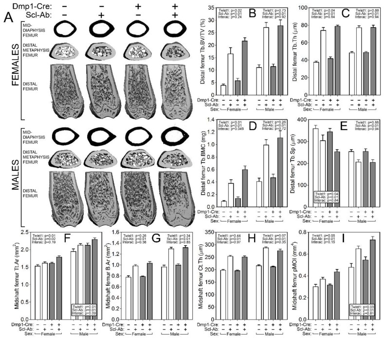
Figure 3.
µCT-derived measurement of the distal femur metaphyseal cancellous bone and mid-femur cortical bone from Cre-negative (white bars) and 10kbDmp1-Cre-positive (grey bars) Twist1f/f mice at 16 wk of age, treated with or without sclerostin antibody (Scl-Ab) for 6 wk prior to sacrifice. (A) Representative 3D reconstructions of (top row) the midshaft femur, (middle row) the distal metaphysis (proximal view), and (bottom row) the caudal half of the distal femur (the ventral half was digitally removed). Female mice are in the top of the panel, males in the bottom. Quantitative differences in (B) femur trabecular bone volume fraction (BV/TV), (C) trabecular thickness (Tb.Th), (D) trabecular bone mineral content (Tb.BMC), and (E) trabecular separation (Tb.Sp) are shown for female (left side of each panel) and male (right side of each panel) mice. Quantitative differences in (F) femur cortical bone total area (Tt.Ar), (G) bone area (B.Ar), (H) cortical thickness (Ct.Th), and (I) polar moment of inertia (pMOI) are shown for female (left side of each panel) and male (right side of each panel) mice. Data were tested for significance using two-way ANOVA within sex, with Twist1 status and antibody as the main effects (plus interaction), which are reported in the corner or base of each sex grouping; n = 8–11/group.

