Figure 1.
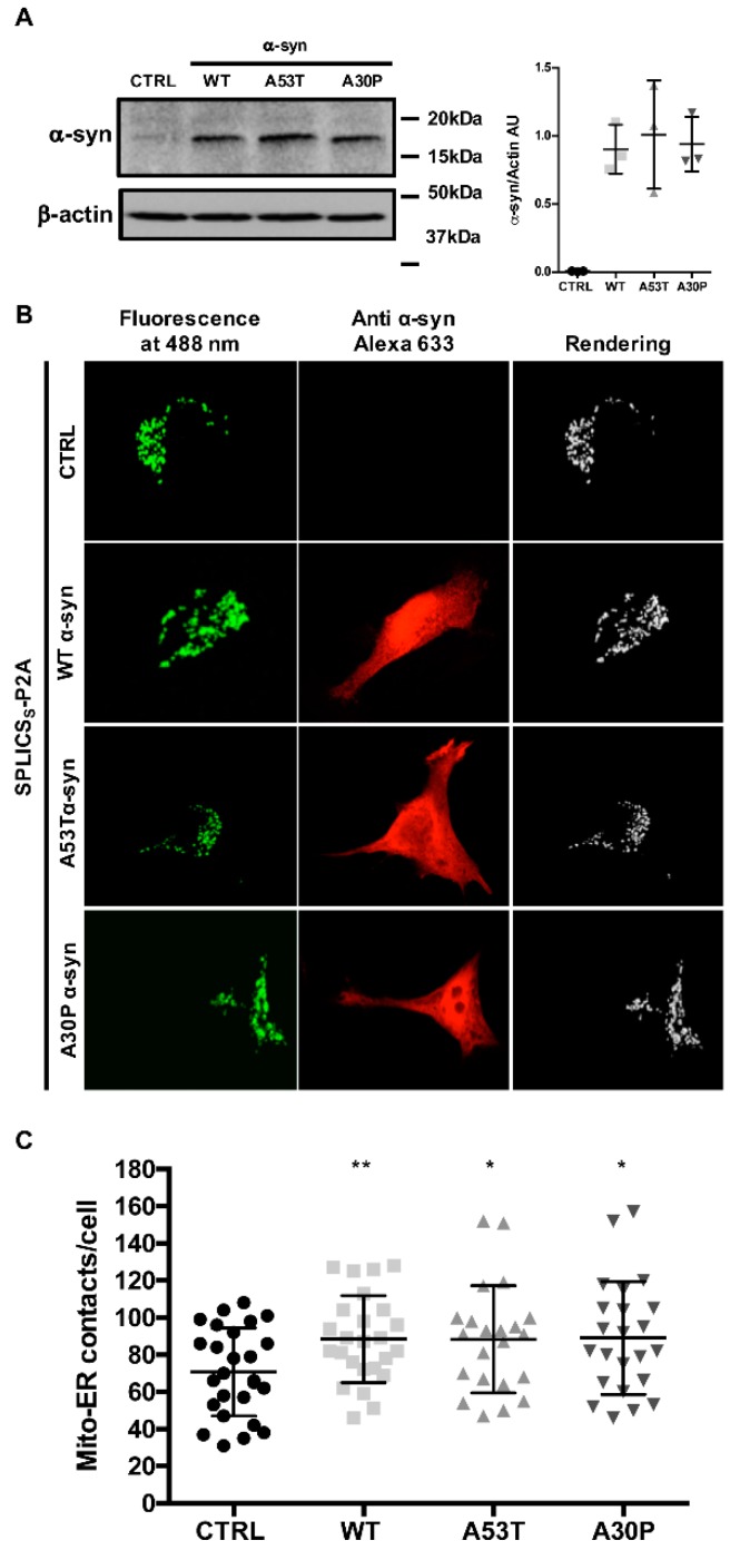
α-syn A53T and A30P mutants physically modulate ER-mitochondria contact sites. (A) HeLa cells were transfected with wt, A53T, and A30P α-syn expression plasmids and analyzed by Western blotting with an anti α-syn antibody. Equal loading was verified by probing the membrane with an anti β-actin antibody. Quantification of three independent experiments (B) HeLa cells were co-transfected with wt, A53T and A30P α-syn expression vectors and the SPLICSS sensor to assess short-range ER-mitochondria associations. Reconstitution of the fluorescent signal was observed upon 488 nm wavelength excitation in α-syn positive cells probed with an anti α-syn primary antibody and revealed by an Alexa 633 secondary antibody. The 3D rendering of the Z-stacks acquired for the SPLICSS probe is shown on the right. (C) Quantification of the ER-mitochondria contact sites/cell in the different conditions is shown as mean ± SD. *, p < 0.05, **, p < 0.01. One-way ANOVA test retrieved a p-value of 0.04. Unpaired Student’s two-tailed t-test was used for two-group comparison. No correction was applied since the same SD was assumed. Dunnet’s post-test was also applied to compare wt and α-syn mutants each other but no significance was detected. The asterisks refer to Student’s two-tailed t-test where the comparison was for each independent sample vs. mock cells.

