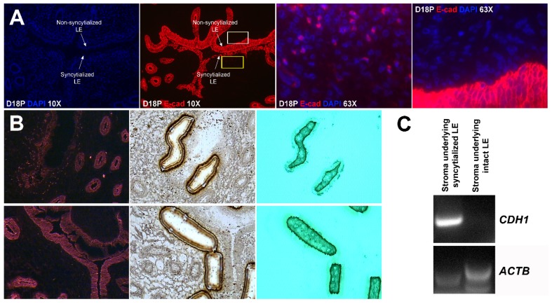
Figure 3.
Localization of E-cadherin-positive cells within the uterine stroma during active syncytialization at implantation sites of sheep. (A) Immunofluorescence staining for E-cadherin (E-cad, red) in the endometrium on day 18 of pregnancy. The third panel represents the region indicated by the yellow box in the second panel. The fourth panel represents the region indicated by the white box in the second panel. E-cadherin-positive cells are present within the stroma underlying actively syncytializing uterine LE. The width of field for the microscopic image captured at 10× and 63× is 890 and 140 μm, respectively. (B) Laser capture microdissection (LCM) used to collect cells within the stroma underlying syncytializing LE (top row) and cells within the stroma underlying intact uterine LE (bottom row). Frozen endometrial tissue sections on day 18 of pregnancy stained with E-cadherin immediately before microdissection (top and bottom rows, first panels). The same tissue section is shown with the missing cells after microdissection (top and bottom rows, middle panels). The LCM cells attached to the CapSure Macro LCM Caps (top and bottom rows, third panels). The width of field for the microscopic image is 631 μm. (C) RT-PCR analysis of CDH1 mRNA in the stromal cells captured by LCM. ACTB was used as a positive control. CDH1 mRNA was detected in the LCM cells isolated from stroma underlying actively syncytializing LE.

