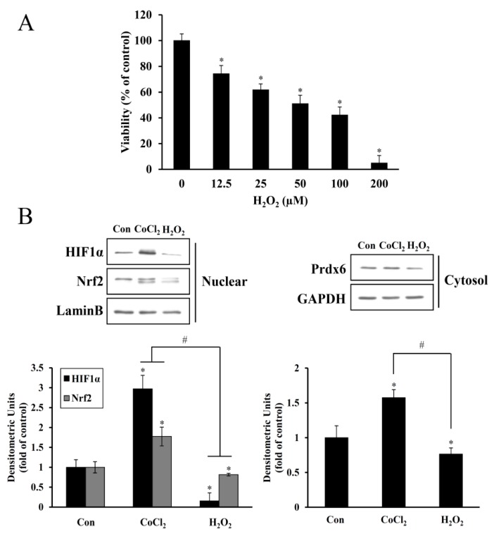
Figure 3.
Inhibitory effect of H2O2 on HEI-OC1 cell viability and HIF-1α, NRF-2, and Prdx6 expression. (A) After exposure to 0–200 μM H2O2 for 24 h, cell viability was determined using the CCK-8 assay. Data are presented as means ± SDs for the three independent experiments, expressed as a percentage of the untreated control. * p < 0.05, compared with the untreated control. (B) Cells treated with 200 μM CoCl2 for 6 h or 100 μM H2O2 for 24 h were harvested, and nuclear and cytosolic proteins were analyzed by immunoblotting for HIF-1α, Nrf-2, and Prdx6. Protein bands were quantified using densitometry, and their abundances were expressed relative to the density of Lamin B or GAPDH band. The ratio of HIF-1α and Nrf-2 to Lamin B or Prdx6 to GAPDH are presented as fold changes relative to the untreated control. Data are presented as the means ± SDs of three independent experiments (* p < 0.05, compared with the control; # p < 0.05, CoCl2 versus H2O2).

