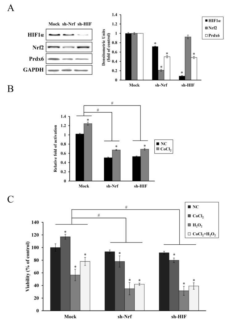
Figure 5.
Inhibitory effect of Nrf-2 and HIF-1α knockdown on Prdx6 expression and preconditioning induced by CoCl2. (A) HEI-OC1 cells transfected with the pLKO.1(mock), pLKO-mNrf-2 (sh-Nrf), or pLKO-mHIF-1α (sh-HIF) were treated with 200 μM CoCl2 for 6 h. Next, Nrf-2, HIF-1α, and Prdx6 expression were analyzed via immunoblotting. Individual bands were quantified densitometrically and normalized to GAPDH. Values in the graph are presented as fold changes relative to the mock transfectant, expressed as means ± SDs of three independent experiments (* p < 0.05, compared with the mock control) (B) Cells were cotransfected with the pGL3-mPx luciferase reporter vector and each KD plus the β-galactosidase plasmid. After 24 h, cells were treated with 200 μM CoCl2 for 6 h, and luciferase activities were determined. Luciferase activities were normalized to that of β-galactosidase. Data are presented as means ± SDs for three independent experiments, expressed as a fold change relative to the mock transfectant. * p < 0.05, compared with the untreated control (NC), # p < 0.05, mock group versus sh-Nrf or sh-HIF group. (C) Cells transfected with mock or each KD plasmid were treated with 200 μM CoCl2 for 6 h, 25 μM H2O2 for 9 h, or CoCl2 plus H2O2, and cell viability was evaluated using the CCK-8 assay. Data are expressed as a percentage of the mock/untreated control and presented as means ± SDs for three independent experiments. * p < 0.05, compared with the untreated control (NC), # p < 0.05, mock group versus sh-Nrf or sh-HIF group.

