Figure 7.
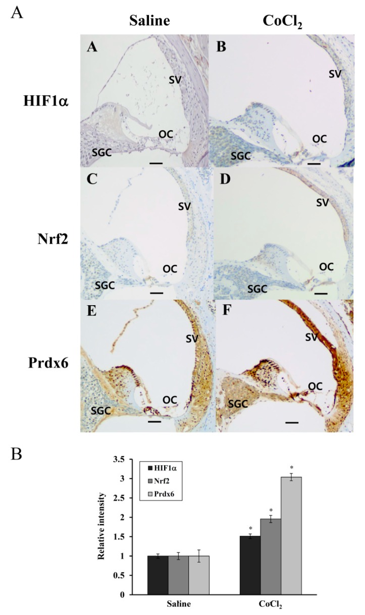
HIF-1α, Nrf-2, and Prdx6 expression in the cochleae of saline- or CoCl2-injected mice. Mouse cochlear paraffin sections were used for immunohistochemical analyses of HIF-1α, Nrf-2, and Prdx6 expression. (A) The expressions of HIF-1α (A: saline-injected, B: CoCl2-injected), Nrf-2 (C: saline-injected, D: CoCl2-injected), and Prdx6 (E: saline-injected, F: CoCl2-injected) are marked with brown-colored deposits and nuclei are counterstained with hematoxylin. OC, organ of Corti, SV, stria vascularis; SGC, spiral ganglion cells. Original magnification ×100, scale bar = 100 μm. (B) The intensities of the dark brown dots in images were quantified using an Image J program. Data in the graph is presented as fold changes relative to the saline-injected cochleae, expressed as ± SDs of three independent experiments. * p < 0.05, compared with the saline-injected cochleae.

