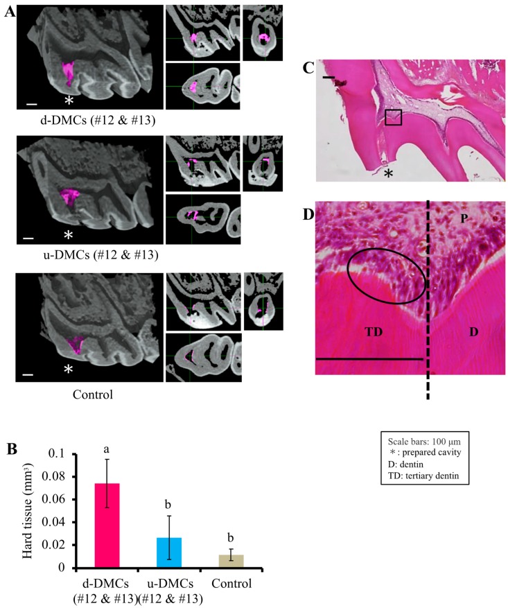
Figure 2.
Effects of the fractions on tertiary dentinogenesis. (A) µ-CT images of tertiary dentin (pink area) induced using fractions #12 and #13 from d-DMCs or u-DMCs, or the control as pulp capping materials. (B) Quantification of the tertiary dentin volume (n = 3 per group). (C,D) Panoramic and high-power H-E-stained images of tertiary dentin formation promoted by d-DMC fractions. An odontoblast-like cell layer and tubular-like structure were observed in the circled area in (D) (D = primary dentin, TD = tertiary dentin, and P = pulp). Images are representative of three independent experiments. Quantitative data are means ± s.e.m. Groups with similar lowercase letters (i.e., a and b) are not significantly different (p > 0.05).

