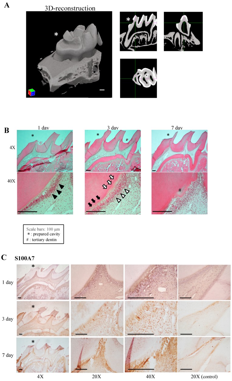
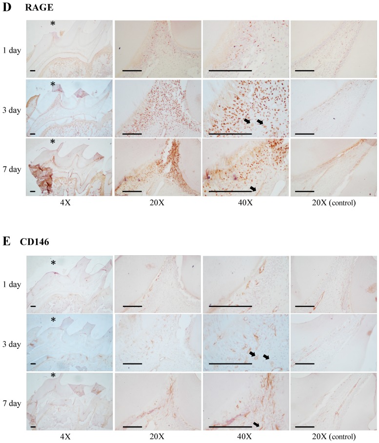
Figure 6.
Localization of protein S100-A7-related proteins in the wound healing process. (A) Three-dimensional reconstruction of a cavity-prepared tooth. (B) Panoramic and high-power H-E-stained images of pulp tissue after cavity preparation. Detachment or disappearance of odontoblasts is indicated by black arrow heads and reattachment of newly differentiated odontoblast-like cells is indicated by white arrow heads. Black arrows indicate the ‘normal’ predentin area, and white arrows indicate the cavity affected predentin-like area. Immunohistochemical staining of S100-A7 (C), receptor for advanced glycation end-products (RAGE) (D), and CD146 (E). Pulp horn tissue of the non-cavity side was used as an internal control. Dual-positive cells for RAGE and CD146 are indicated by black arrows (D,E). Scale bar: 100 μm (* = prepared cavity; # = tertiary dentin). Images are representative of three independent experiments.


