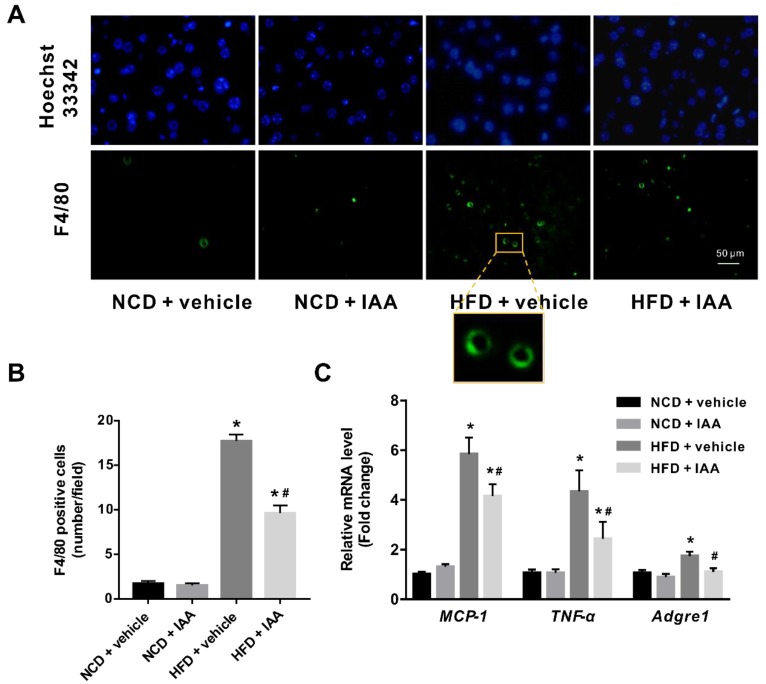
Figure 6.
Inflammatory stress in liver of mice induced by high-fat diet (HFD) was ameliorated by indole-3-acetic acid (IAA) treatment. (A) Immunofluorescence analysis for F4/80 antigen in liver tissue section. Scale bar represents 50 µm. (B) Count of F4/80 positive cells per field (at least six field per mice) (C) Relative mRNA abundance of MCP-1, TNF-α, and Adgre1. Values are presented as the mean ± standard error of the mean. n = 6. * p < 0.05 vs. NCD + vehicle; # p < 0.05 vs. HFD + vehicle.

