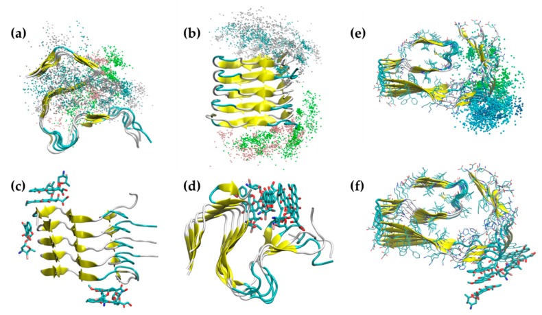
Figure 6.
Iododoxorubicin binding to (a–d) 2MXU and (e,f) 5OQV fibril structures. Superposition of iododoxorubicin coordinates (center of mass) during the 2MXU MD simulations (a,b) and snapshots of the main binding regions (c,d). Superposition of IDOX coordinates during the 5OQV MD simulation (d) and a snapshot of the main binding area (f).

