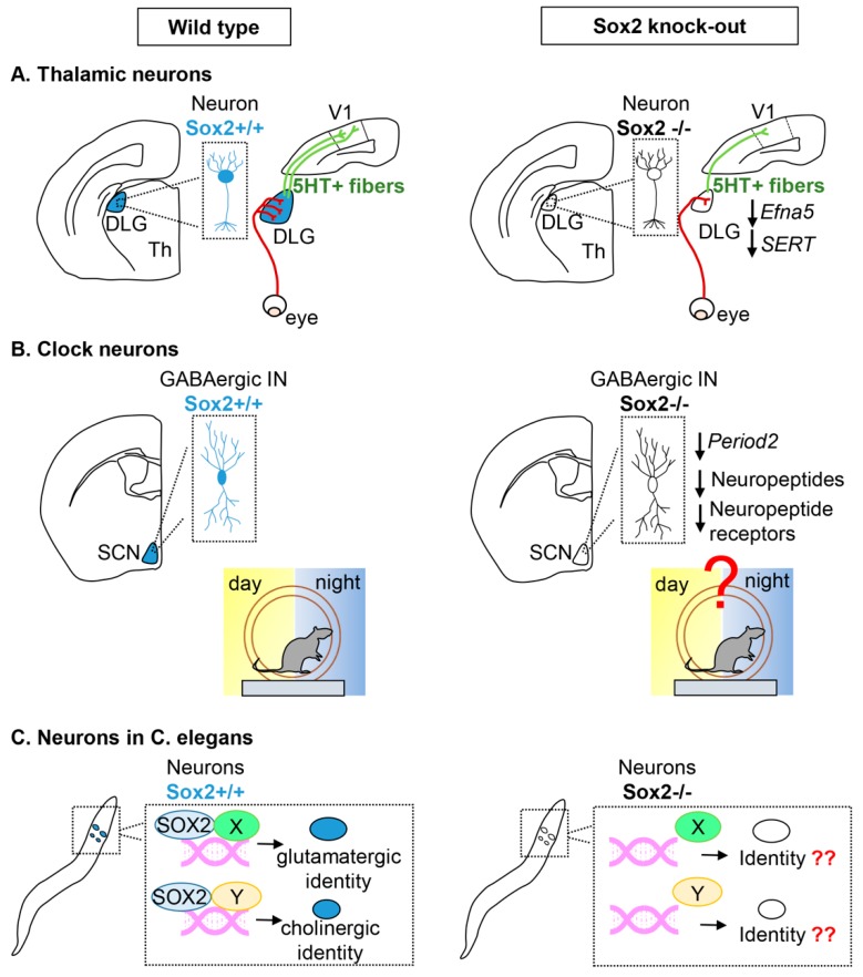
Figure 2.
Differentiated neuronal cells requiring Sox2 function. Neuronal cells expressing Sox2 in wild type (left) are colored in blue. The corresponding cell types in Sox2 knock-out mice (right) are uncolored. (A) Thalamic neurons within the mouse sensory thalamic nuclei. In wild type animals, axons outgrowing from the retina reach the DLG in a spatially ordered pattern; DLG neurons develop 5HT-positive projections to the primary visual cortical area (V1). In mutant animals, the DLG is reduced, and the eye-DLG projections are reduced and disorganized; the DLG-V1 projections are also defective. A reduction of the expression of Efna5 and SERT in the mutant contributes to the defects. Abbreviations: DLG, dorsolateral geniculate nucleus; Th, thalamus; V1, primary visual area of the cerebral cortex; 5HT, 5-hydroxytryptamine, i.e., serotonin. (B) Clock neurons within the mouse suprachiasmatic nucleus. In wild types, Sox2-expressing GABAergic interneurons in the SCN are essential for establishing circadian rhythms. In mutants, circadian rhythms are perturbed; a decrease in the expression of Period2, and of neuropeptides and their receptors contribute to the defect. Abbreviations: SCN, suprachiasmatic nucleus; IN, interneurons. (C) Neurons of C. elegans. In wild types, Sox2 is expressed in specific neuronal cell types, both glutamatergic and cholinergic, and binds to DNA with different partners (X, Y). In mutants, the terminal differentiation of these neuron types is defective.

