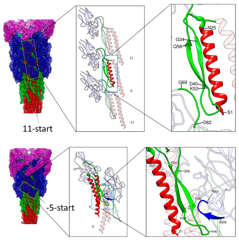
Figure 3.
Intermolecular interactions along the 11-start and −5-start direction. Left panels are side views of the hook with the four domains of FlgE colored as in Figure 1, with outer domains removed towards the bottom. The yellow boxes in the left panel indicate the arrays of FlgE subunits magnified in the middle panel: Three subunits in the 11-start helical line in the upper row and two subunits in the −5-start in the lower row. Magnified images of some parts of FlgE are also colored as in Figure 1 in the middle and right panels. In the upper row, the N-terminal helix and the long β-hairpin of domain Dc of subunit 0 are colored red and green, respectively. In the lower row, the N- and C-terminal helices and the long β-hairpin of subunit 0 are colored red and green, respectively, and the tip loop of a short β-hairpin at the bottom of domain D1 of subunit −5 is colored blue. These colored parts in the middle panels are highlighted with light-blue boxes to indicate the portions further magnified in the right panels.

