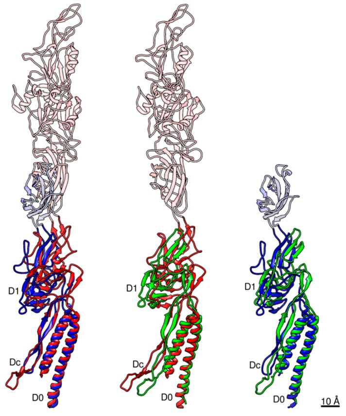
Figure 5.
Pairwise structural comparison between Salmonella FlgE and FlgG and Campylobacter FlgE. Salmonella FlgE and Campylobacter FlgE are compared in the left panel, Salmonella FlgG and Campylobacter FlgE in the middle, and Salmonella FlgE and FlgG in the right. The Cα ribbon models are colored only for domains D0-Dc and D1, with Salmonella FlgE in blue, FlgG in green and Campylobacter FlgE in red. Domain D0 is used to superpose these molecules. The relative arrangements of domains D1 and D0-Dc between these three molecules are slightly different to one another as shown in this figure. The extra part of domain D1 present in FlgE and missing in FlgG is the triangular loop involved in the intersubunit sliding interactions between D1 and D2 along the protofilament for its compression and extension.

