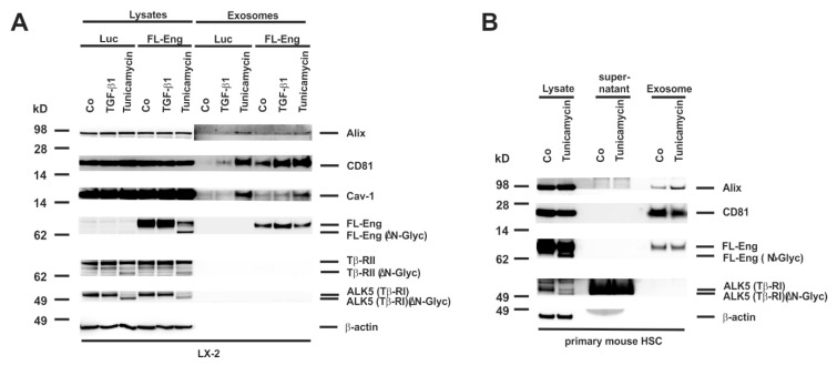
Figure 8.
Isolation of exosomes from HSC treated with tunicamycin. (A) Cell extracts or exosome fractions prepared from LX-2 which were infected with FL-Eng or a control virus expressing luciferase (Luc). Subsequently, the cells were treated with TGF-β1 or an inhibitor of N-glycosylation (tunicamycin) and analysed by Western blot for expression of indicated proteins. Please note, that only properly glycosylated Eng is directed to the exosomal compartment. (B) Cell protein extracts, supernatants, and exosomes were prepared from primary mouse HSC and analysed for the presence of indicated proteins demonstrating that endogenous Eng is only directed to exosomes when properly glycosylated.

