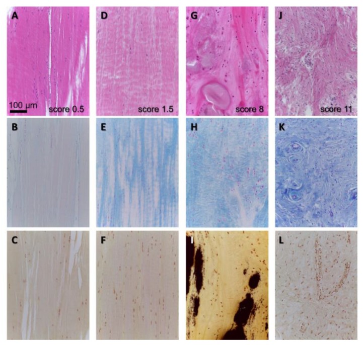
Figure 4.
Histopathological features of human ACL degeneration in OA. (A,D,G,J): HE staining to give an overview over tissue organization. (B,E,H,K): Alcian blue staining to visualize distribution of sulfated glycosaminoglycans (blue), (C,F,I,L): van Kossa staining to detect calcium deposits (black). The scoring results with the scoring system used by [11] are depicted in (A,D,G,J), (A–C): nearly unchanged, (G–I): chondroid metaplasia and focal calcification, (J–L): ECM disintegration, hypercellularity, hypervascularization.

