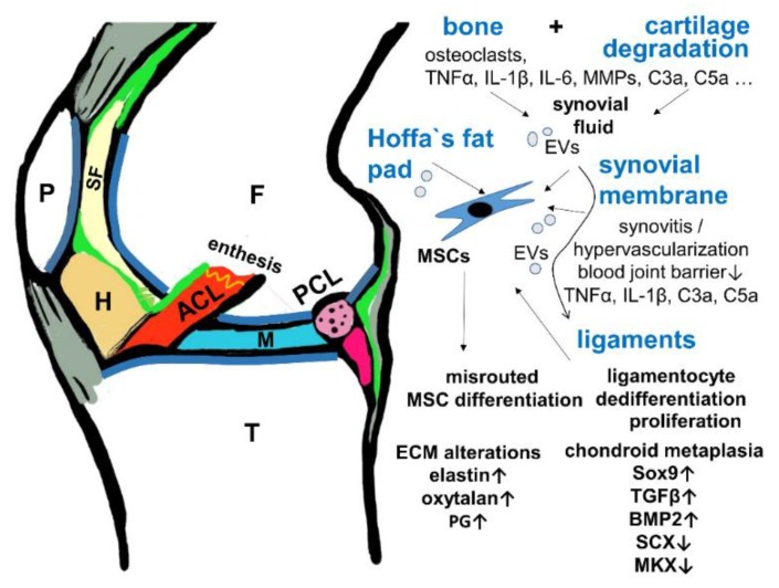
Figure 6.
Hypothesis of the cross-talk between cartilage/bone and ligaments in OA. A scheme of a sagittal section of the knee is shown depicting the interrelation of the joint tissues involved in OA. ACL: red, F: femur, H: Hoffa’s fad pad, brown, M: meniscus, P: patella, PCL: transected, pink, SF: synovial fluid, pale yellow, T: tibia, green: synovial membrane, gray: patellar tendon, ligament and joint capsule. BMP2: Bone morphogenetic protein, EV: extracellular vesicles, PG: proteoglycans, MKX: Mohawk, SCX: scleraxis, TGFβ: transforming growth factor β.

