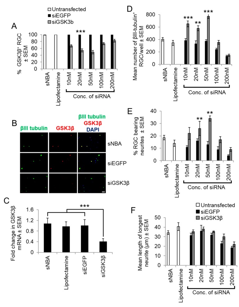
Figure 4.
siGSK3β-mediated knockdown in retinal cultures prepared from adult rats at five days after ONC. (A) Quantification of the proportion of GSK3β+ RGC with increasing concentrations of siGSK3β when compared to sNBA and Lipofectamine 2000-treated controls. (B) Representative retinal cultures established five days after ONC in vivo to activate retinal glia and treated with siGSK3β (50 nM) demonstrated a lack of GSK3β (red) detection in βIII-tubulin+ RGC (green), whilst abundant immunoreactivity was present in βIII-tubulin+ RGC in sNBA and siEGFP-treated (50 nM) control cultures. (Scale bar in B = 20 µm; ** = p < 0.01; *** = p < 0.001). (C) Analysis of GSK3β mRNA levels in cultured adult rat retinal cells after transfection with the optimal concentration of siGSK3β (50 nM) to confirm GSK3β knockdown. (*** = p < 0.001). (D) Quantification of the proportion of surviving βIII-tubulin+ RGC with increasing concentrations of siGSK3β shows that 50 nM optimally promotes RGC survival. (E) Quantification of the proportion of RGC bearing neurites with increasing concentrations of siGSK3β shows that 50 nM was optimal to stimulate initiation of neurite outgrowth. (F) Quantification of the longest RGC neurite length shows that increasing concentrations of siGSK3β does not affect the length. (** = p < 0.01; *** = p < 0.001). n = 2 wells/treatment, 3 independent repeats (total n = 6 wells/treatment).

