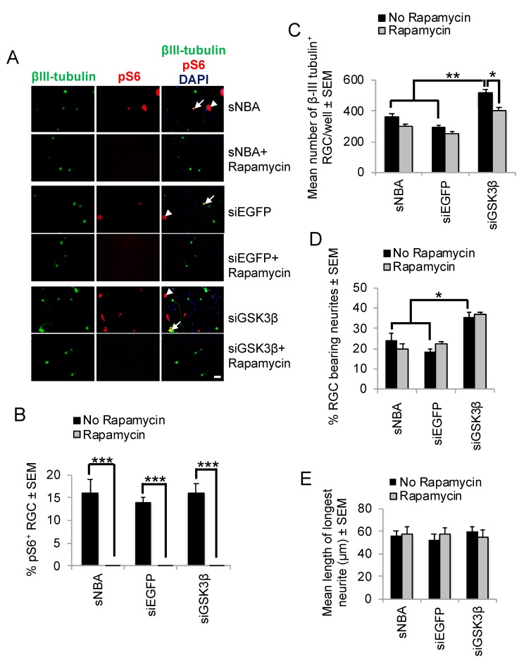
Figure 5.
Effects of Rapamycin on siGSK3β-mediated RGC survival and mTORC1 activity in vitro. (A) Representative retinal cultures established five days after ONC in vivo to activate retinal glia, treated for three days with sNBA, siEGFP, and siGSK3β in the presence and absence of the mTORC1 inhibitor Rapamycin and immunostained for βIII-tubulin (green) and pS6 (red), with DAPI (blue) as a nuclear counterstain. Note that pS6 is detected both in βIII-tubulin+ RGC (long arrows) and βIII-tubulin− cells (short arrows) and the abolition of pS6 immunostaining in the presence of Rapamycin. (B) The % of RGC exhibiting pS6 immunoreactivity indicated abolition of pS6 expression in the presence of Rapamycin, but there was no significant effect of siGSK3β on the proportion of pS6+ RGC (* = p < 0.05, ** = p < 0.01, *** = p < 0.001). (C) Quantification of the number surviving βIII-tubulin+ RGC after treatment with siGSK3β in the presence of Rapamycin shows that RGC survival is significantly enhanced. (Scale bar in A = 20 μm; ** = p < 0.01). (D) Quantification of the % RGC bearing neurites and (E) length of the longest RGC neurite are both significantly enhanced after siGSK3β treatment, but this effect was unaffected by Rapamycin. n = 2 wells/treatment, 3 independent repeats (total n = 6 wells/treatment).

