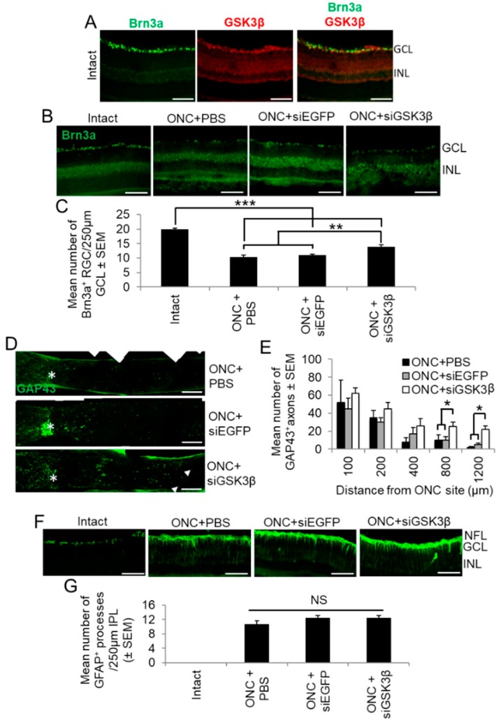
Figure 7.
Cellular localisation of GSK3β and the effects of siGSK3β on RGC neuroprotection in vivo. (A) Retinal sections from uninjured adult rats immunostained for Brn3a (green) and GSK3β (red), demonstrating that constitutive expression of GSK3β was limited to the GCL, where it localised to Brn3a+ RGC (INL = inner nuclear layer). (B) siGSK3β protects RGC (Brn3a+) from death at 24 days after ONC, compared to ONC + PBS and ONC + siEGFP. Intact uninjured controls show baseline levels of Brn3a+ RGC. (C) Quantification of Brn3a+ RGC survival in the 250 µm counting area of the GCL. (Scale bars in A and B = 100 µm; ** = p < 0.01; *** = p < 0.001). (D) Longitudinal ON sections immunostained to demonstrate GAP43+ regenerating axons (arrows) after ONC + PBS (top panel), ONC + siEGFP (middle panel) and ONC + siGSK3β (bottom panel). The asterisk demarcates the ON site and the boxed area in the lower panel represents the magnified area. (E) Quantification of GAP43+ regenerating axons 100, 200, 400, 800, and 1200 μm beyond the ONC site in eyes after intravitreal injection of PBS, siEGFP, and siGSK3β. (Scale bars = 200 μm; * p < 0.05). (F) GFAP+ glial activation occurs after ONC and is not further enhanced by siGSK3β treatment. (G) Quantification of the number of GFAP+ glial processes crossing a 250 µm line in the inner plexiform layer corroborates this observation. (Scale bar in F = 100 µm; NS = not significant). n = 6 eyes (from 6 rats)/treatment.

