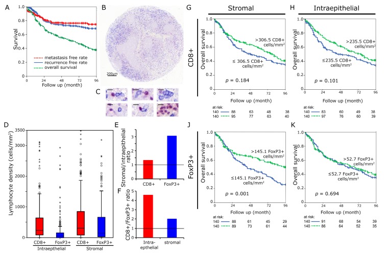
Figure 1.
CD8+ and FoxP3+ cell densities in head and neck cancer. Kaplan Meier plots for metastasis free rate, recurrence free rate and overall survival in the complete cohort of 280 patients (A). Tissue samples were processed into tissue microarrays using a core diameter of 2 mm. Scale bar 200 µm (B). High power views (1:400) with immunohistochemical double staining for FoxP3 (red nucleic staining) and CD8 (blue predominantly membranous staining). (Scale bars 10 µm) (C). Lymphocyte densities (cells/mm²) in the intraepithelial and stromal compartment were separately analyzed (D). Stromal/intraepithelial ratio of CD8+ and FoxP3+ cells (E). CD8+/FoxP3+ ratio in intraepithelial and stromal compartment (F). Kaplan Meier plots for densities of CD8+ lymphocytes in the stromal (G) and intraepithelial H as well as FoxP3+ cells in the stromal (J) and epithelial (K) compartment (overall survival). Cut-off values were the median.

