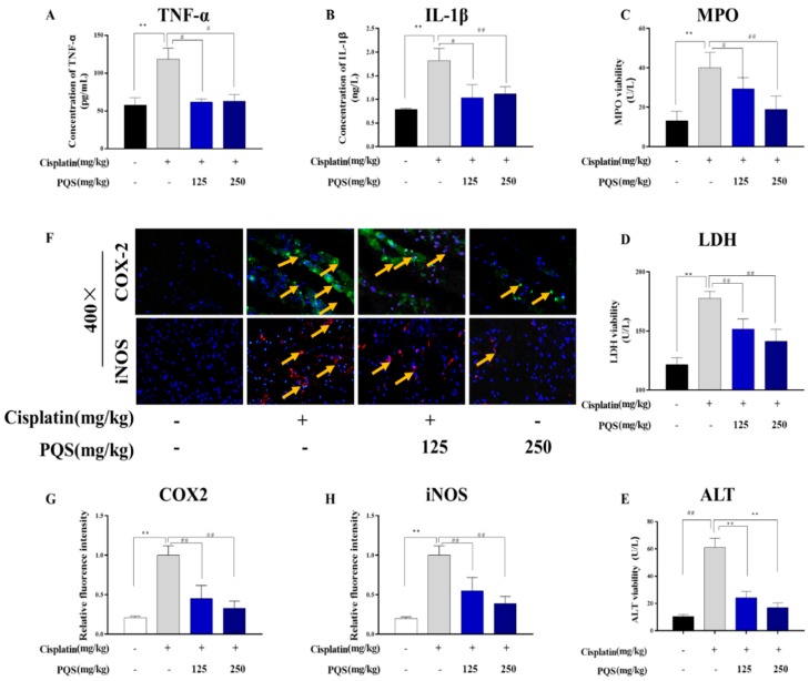
Figure 4.
Effect of PQS on cisplatin-induced changes in inflammatory markers in heart tissues of mice. (A) Tumor necrosis factor-a (TNF-α); (B) Interleukin-1β (IL-1β); (C) Myeloperoxidase (MPO) activity; (D) Lactate dehydrogenase (LDH); (E) ALT activity; (F) PQS exerted great changes on expression of COX-2 and iNOS in heart tissues, the expression levels of COX2 (Green) and iNOS (Red) in tissue section isolated from different groups were assessed by immunofluorescence. (G) Quantitative analysis of scanning densitometry for cleaved COX-2 (H) Quantitative analysis of scanning densitometry for cleaved iNOS. All data were expressed as mean ± S.D. ** p < 0.01 comparing with normal group. # p < 0.05 or ## p < 0.01 comparing with cisplatin group.

