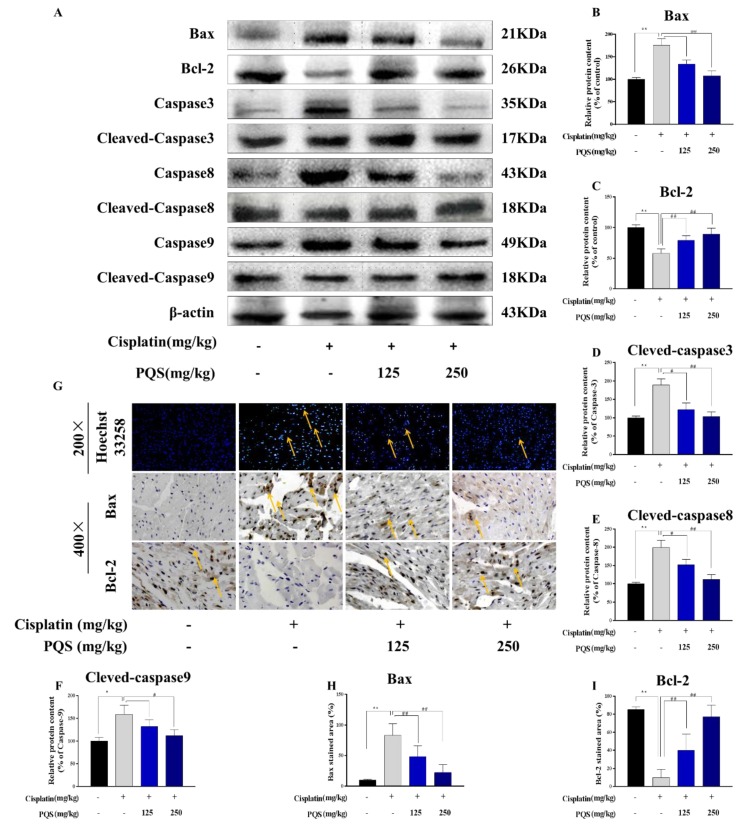
Figure 7.
(A) The blots of Bax, Bcl-2, Bad, caspase 3, cleaved caspase 3, caspase 8, cleaved caspase 8 and caspase 9, cleaved caspase 9 were standardized to that of β-actin; Quantitative analysis of scanning densitometry for Bax (B), Bcl-2 (C), Caspase-3 (D), Caspase-8 (E), Caspase-9 (F). (G) Representative photomicrographs of cardiac immunohistochemically staining for Hoechst 33258, (H) Bax staining area. The percentage of apoptosis (I) and Bcl-2 staining area in indicated groups. Scale bars data were expressed as mean ± S.D. * p < 0.05 or ** p < 0.01 comparing with normal group. # p < 0.05 or ## p < 0.01 comparing with cisplatin group.

