Figure 2.
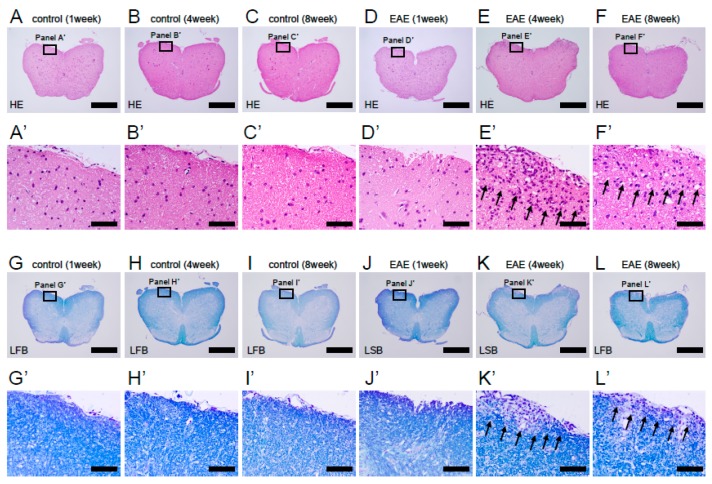
H&E (A–F, A’–F’) and LFB staining (G–L, G’–L’) of lumbar spinal cord sections obtained from control (A–C, A’–C’, G–I, and G’–I’) and MOG-immunized mice (D–F, D’–F’, J–L, and J’–L’) at 1, 4, and 8 weeks after treatment. Infiltration of inflammatory cells and significant demyelination was observed 4 and 8 weeks after treatment in EAE mice, whereas no demyelination was observed at any time points in control mice. Results displayed are representative of three replicates (N = 3). Scale bars = 500 µm (A–L) and 50 µm (A’–L’). Abbreviations: H&E, hematoxylin and eosin; LFB, luxol fast blue; MOG, myelin oligodendrocyte glycoprotein; EAE, experimental autoimmune encephalomyelitis.

